

Dental X-rays help your dentist assess structures that cannot be seen during a visual examination, such as tooth roots and the surrounding bone. Different types of images support the assessment of decay, gum disease and jaw conditions, as well as planning for dental implants and root canal treatment.
Our imaging facilities are located at ProDent, so examinations available here can be carried out without travelling to a separate imaging centre. Your dentist decides which images are needed following a consultation and examination.
What are dental X-rays used for?
- Assessing tooth decay, including areas between teeth and around fillings that are difficult to inspect directly.
- Assessing gum disease, particularly changes in the bone supporting the teeth.
- Planning orthodontic treatment, using panoramic and cephalometric images where indicated.
- Planning implant treatment, using CBCT where needed to assess bone dimensions and nearby anatomical structures.
- Investigating abnormalities, such as inflammatory changes, cysts, injuries or changes in the jawbone.
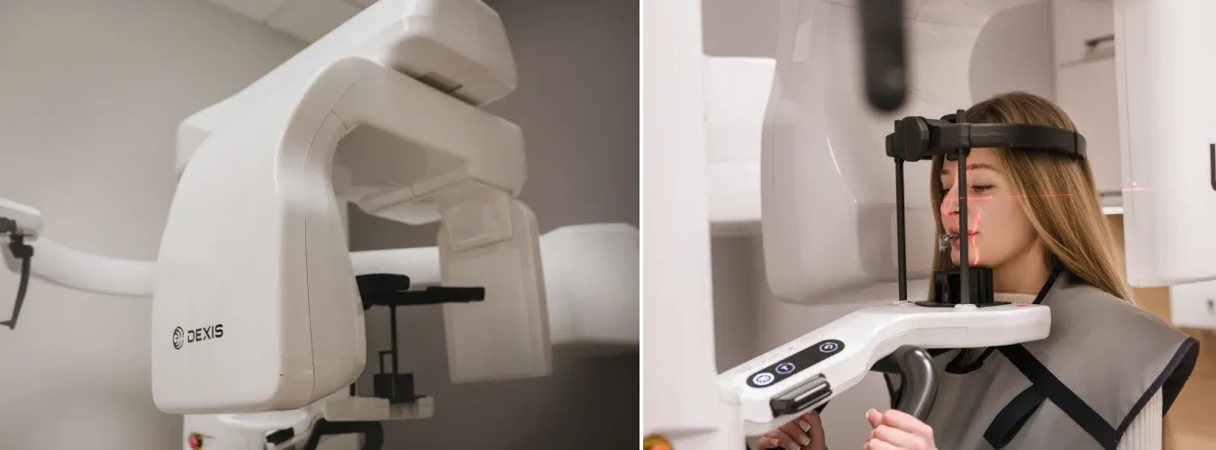
DEXIS OP 3D LX imaging
At ProDent in Nysa, we use the DEXIS OP 3D LX for 2D and 3D imaging. The area scanned is selected to address a specific clinical question, from a focused area around a tooth to a larger region of the jaws.
The field of view and imaging settings are chosen for the examination. A larger scan is not automatically more useful: the aim is to obtain the information needed to plan care while limiting unnecessary exposure.
Explore the other devices used at our clinic on our equipment and technology page.
Types of dental imaging
Intraoral X-rays
These images are taken with the image receptor positioned inside the mouth.
| Examination | What it shows |
|---|---|
| Periapical X-ray | A tooth from the crown to the root tip, together with the surrounding bone. |
| Bitewing X-ray | The crowns of upper and lower teeth and the areas between them. |
| Occlusal X-ray | A wider area of the mouth, including part of the dental arch. |
Extraoral X-rays
- Panoramic X-ray (OPG): an overview of the teeth and jaws in a single image.
- Cephalometric X-ray: a side view of the skull, often used when planning orthodontic treatment.
- Temporomandibular joint imaging: images of the jaw joints, selected according to the clinical problem.
3D CBCT scans
Cone beam computed tomography (CBCT) produces three-dimensional images of the teeth and jaws. Your dentist may recommend it when conventional 2D images do not provide enough information, for example in implant planning, oral surgery or selected complex root canal cases.
Not every treatment requires a CBCT scan. The choice depends on what needs to be assessed in your individual case.
How imaging supports treatment
Images can help identify changes before symptoms develop, show the anatomy relevant to a procedure and provide a basis for comparing findings during follow-up. They form part of the assessment alongside your symptoms, dental history and clinical examination.
No single image answers every diagnostic question. Your dentist will explain why a particular examination is recommended.
Radiation exposure and safety
Dental X-rays use ionising radiation, so an examination should have a clinical reason. The dose depends on the type of image, equipment, settings and area scanned. A 2D X-ray and a CBCT scan do not have the same exposure.
We select the examination and imaging area according to the information needed and follow the radiation protection procedures applicable to the examination. Previous images may help avoid unnecessary repeat examinations, so tell your dentist if you have them.
Further information: American Dental Association guidance on dental X-rays.
Your imaging appointment at ProDent
We provide dental 2D imaging and CBCT at Prudnicka 3/1u, Nysa, Poland, to support diagnosis and treatment planning. Images taken as part of your care at ProDent are available to your treating dentist.
You can find indicative prices for panoramic X-rays, cephalometric imaging and CBCT in our diagnostic imaging price list. The examination required is confirmed for your case.
Preparing for your examination
Before attending, ask reception about referral requirements and the examination requested. Tell the team if you are pregnant or may be pregnant, and bring previous images if you have them. Fasting is usually unnecessary. Follow the staff’s instructions about removing metal items around your head.
Having treatment at another practice?
Contact reception to confirm whether we can carry out the requested examination and how the images can be provided to your dentist.